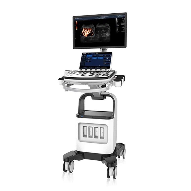
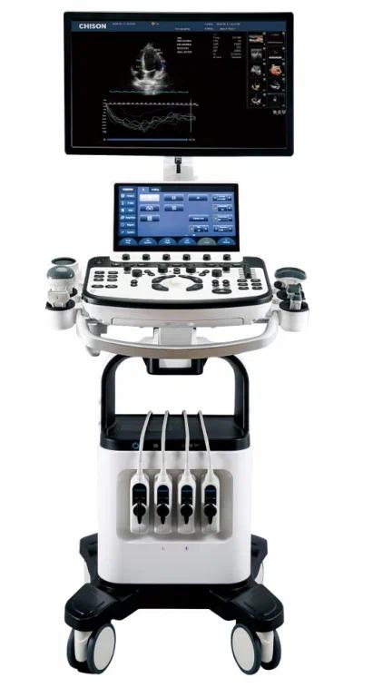
Chison Qbit 12 Ultrasound Unit
Price:
- on request
Specifications:
- Active probe ports - 3 + 1 additional
- Modes - CW, Color Doppler, real-time 3D imaging, 3D imaging, Stress Echo, Compound Imaging, contrast imaging, SonoContrast, Color Panoramic Imaging
- Dimensions - 55 x 45 x 145 cm
- Weight - 60 kg
- Country of origin - China
- Medical device type code - 260250
Description
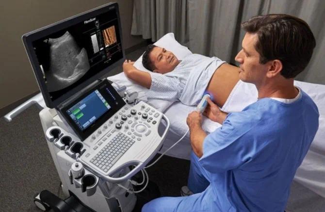
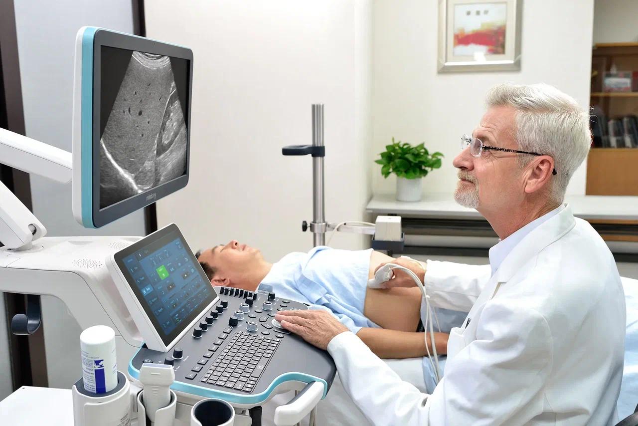
Stationary ultrasound scanners are medical equipment designed for ultrasound examination of internal organs and tissues. They are used in the diagnostic departments of hospitals, clinics, and medical centers to detect various conditions.
The ultrasound scanner generates high-frequency sound waves that penetrate the body. These waves reflect off the boundaries between different tissues and organs, creating an echo. The scanner picks up this echo and converts it into an image on the monitor screen.
Ultrasound scanners are used to diagnose various conditions, including tumors, cysts, stones, inflammation, developmental abnormalities, and more. They allow a physician to assess the size, shape, structure, and function of organs and tissues, and to detect pathological changes.
Ultrasound scanners are a safe, non-invasive diagnostic method. They provide a detailed image of internal organs and tissues, and can be used to monitor treatment and evaluate its effectiveness.
Key Advantages
- - Chison Qbit 12 offers numerous scanning modes, including continuous-wave Doppler (CW), color Doppler, real-time 3D imaging, and more, making it a versatile tool across medical fields;
- - 3D imaging capability provides volumetric images, especially useful in cardiology, obstetrics, and gynecology for more detailed diagnosis;
- - Stress Echo mode is designed for examining the heart under physical exertion, helping detect hidden cardiac pathologies;
- - The use of contrast agents improves tissue contrast resolution, making images clearer and more informative;
- - SonoContrast technology allows visualization of tissue microvascular flow, important for diagnosing vascular diseases;
- - Color Panoramic Imaging - this mode expands the scan area, providing a more complete picture of the examined region, especially useful when examining large vessels.