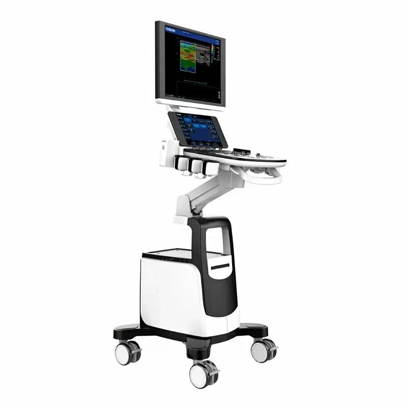
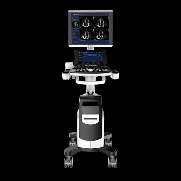
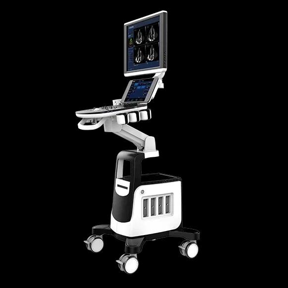
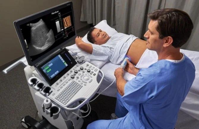
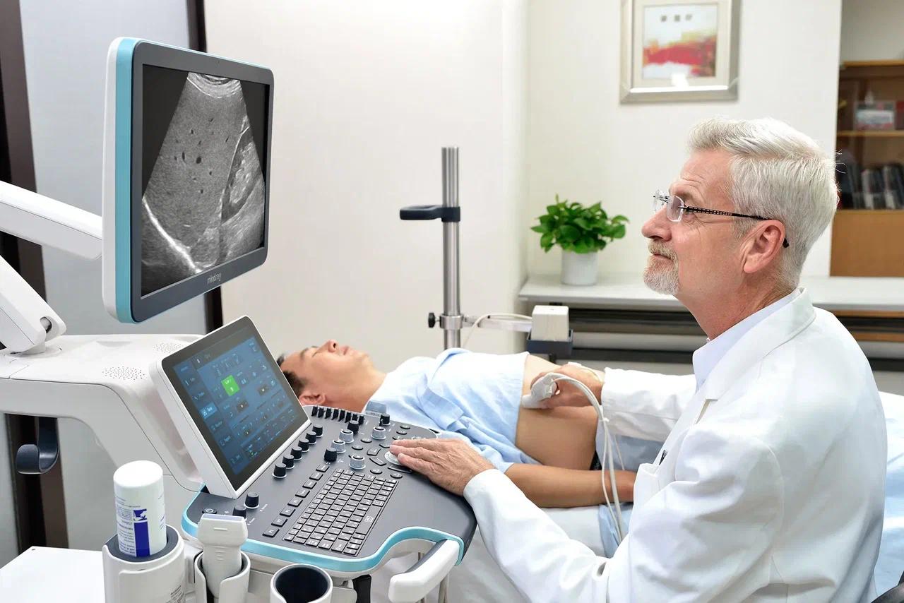
Chison Qbit 10 Ultrasound Unit
Price:
- on request
Specifications:
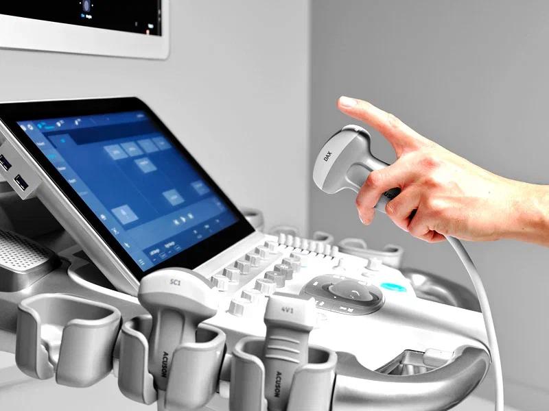
- Active probe ports - 3 + 1 additional
- Modes - B-mode, M-mode, B/M mode, 4D mode, panoramic scanning, trapezoidal mode, strain elastography, stress echocardiography, ECG module support, exam support, automatic image optimization, automatic hemodynamic parameter calculation, multi-beam scanning/compounding
- Dimensions - 55 x 45 x 145 cm
- Weight - 73 kg
- Country of origin - China
- Medical device type code - 260250
Description
Stationary ultrasound scanners are medical equipment designed for ultrasound examination of internal organs and tissues. They are used in the diagnostic departments of hospitals, clinics, and medical centers to detect various conditions.
The ultrasound scanner generates high-frequency sound waves that penetrate the body. These waves reflect off the boundaries between different tissues and organs, creating an echo. The scanner picks up this echo and converts it into an image on the monitor screen.
Ultrasound scanners are used to diagnose various conditions, including tumors, cysts, stones, inflammation, developmental abnormalities, and more. They allow a physician to assess the size, shape, structure, and function of organs and tissues, and to detect pathological changes.
Ultrasound scanners are a safe, non-invasive diagnostic method. They provide a detailed image of internal organs and tissues, and can be used to monitor treatment and evaluate its effectiveness.
Key Advantages
- - High-quality image reproduction thanks to modern design and technology;
- - Versatility and a variety of available probes make it universal equipment for diagnosing various conditions;
- - Affordable pricing compared to similar models makes it an attractive choice for medical facilities;
- - A color Doppler function, essential for examining the vascular system, highlighting blood flow characteristics with color;
- - The ability to examine the smallest details thanks to volumetric 4D imaging;
- - Simultaneous acquisition of up to 3 slices and scanning via a large number of beams, improving image quality and bringing it closer to CT.